Showing posts with label Malformations. Show all posts

Monday, March 14, 2011

Band heterotopia

CASE 1 (7-month-old)


CASE 2 (Pre-adolescent)


Findings

Case 1: Axial and coronal T2-weighted images of the brain in a 7-month-old girl with seizures demonstrate a band of isointense signal within the subcortical white matter, characteristic of band heterotopia.
Case 2: Axial and coronal T1-weighted images demonstrate band heterotopia, better seen in this preadolescent girl due to completion of myelination.


Diagnosis: Band heterotopia


Band heterotopia is a rare neuronal migration anomaly which manifests as homogenous bands of gray matter are interposed between the lateral ventricles and cortical mantle with normal appearing white matter on either side. The overlying cortex may be normal, pachygyric, or display a simplified gyral pattern with short gyri and shallow sulci. At least six morphologically distinct subtypes have been described. Band heterotopias represent a subset of gray matter heterotopia which also includes subependymal and subcortical heterotopia subtypes.

Band heterotopia typically affects female patients as a result of an X-linked dominant inheritance pattern secondary to abnormal function of the doublecortin (DCX) gene (Xp22.3-p23) or less frequently the LIS1 (17p13.3) gene. Male patients can be affected due to sporadic mutations of these genes (41 reported cases in the literature by D’Agostino, et al in 2002). The rate of detectable mutations involving DCX or LIS1 in male patients (42%) is lower than the rate of 85% described in female patients. Dysmorphic features described in patients with band heterotopia include microcephaly (most common), wide nasal bridge, high arched palate, and short stature.

The clinical presentation of band heterotopia can range from normal to nearly normal intelligence and mild developmental delay to frank mental retardation. Seizures are often also present and may begin in the first decade, ranging from partial to generalized or multiple seizure types. The discovery of the underlying brain malformation is due to the onset of seizures in 65% of patients. Eventually 95% of patients with band heterotopias will develop epilepsy. Seizures associated with band heterotopia are often refractory to medical therapy, and surgical therapies such as callosotomy may be performed in these patients. In the series of 30 male patients published in 2002, 46% of patients were refractory to medical therapy and experienced up to 20-30 seizures daily despite trails of multiple therapeutic regimens. Affected male patients tend to have either mild or severe symptoms, whereas, female patients tend to have symptoms within the mild to moderate range of the spectrum from minimal cognitive impairment to severe mental retardation. Posterior involvement, in particular the partial posterior and intermediate posterior subtypes, occur more commonly in male patients. Frontal and diffuse subtypes are more often present in affected female patients.

Friday, December 10, 2010

Capillary Telangiectasia


Findings

There is an ill-defined enhancing focus in the medial right temporal lobe on post gadolinium contrast T1-weighted imaging (Figure 4). There is no corresponding signal abnormality or mass on the precontrast T1-weighted, T2-weighted, or FLAIR images (Figure 1, Figure 2, and Figure 3, respectively). There is no mass effect. On susceptibility-weighted imaging (SWI) the lesion shows hypointensity (Figure 5).


Diagnosis: Capillary Telangiectasia


Brain capillary telangiectasias are benign vascular malformations which are often found incidentally.
They can be visualized by gadolinium contrast and gradient-echo susceptibility or susceptibility weighted imaging, but not through catheter angiography, and may often not be visible on conventional T1/T2, FLAIR, or diffusion-weighted imaging.
Often asymptomatic and usually no treatment is required.

Brain capillary telangiectasias (BCTs) are one of four major types of vascular malformations which occur in the brain (the other three are arteriovenous malformations, cavernous malformations (cavernous angiomas), and developmental venous anomalies (venous angiomas), and represent up to 20% of all intracranial vascular lesions. BCTs consist of multiple ectatic capillaries surrounded by normal brain parenchyma and are usually devoid of calcification, gliosis, extraluminal hemorrhage, and hemosiderin-laden macrophages. BCTs are most common in the midbrain, pons, medulla, and spinal cord, but they are found throughout the central nervous system. Multiple BCTs are possible, especially in certain syndromes (e.g.; ataxia telangiectasia, Osler-Weber-Rendu, or Sturge-Weber syndrome).

Often found incidentally, BCTs are usually benign, small in size, and rarely grow over time. They are rarely symptomatic and are not associated with any particular clinical feature but have been reported to be associated with headache, vertigo, and tinnitus.

BCTs are relatively well visualized through susceptibility weighted imaging where they demonstrate marked signal intensity loss due to deoxyhemoglobin present in slow flowing blood. They are also well visualized through gadolinium-enhanced T1-weighted imaging sequences where they are seen as small faint lesions. BCTs are difficult to visualize through conventional T1/T2, FLAIR, or diffusion-weighted imaging and are considered to be one of the “angiographically occult vascular malformations” due to their small size, tendency to occlude, and sluggish flow.

Friday, November 5, 2010

Pallister-Hall syndrome


60-year-old female with history of a childhood seizure disorder, polydactyly, esophageal narrowing and imperforate anus presents to the ER with acute onset headache.


Findings

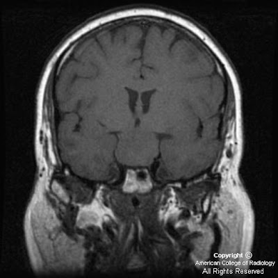
Figure 1: T1 weighted coronal MRI demonstrates an isointense homogeneous suprasellar mass extending from the floor of the third ventricle.
Figure 3: T2 weighted coronal image, again demonstrating a solid homogeneous suprasellar mass with lack of any cystic components or surrounding edema.
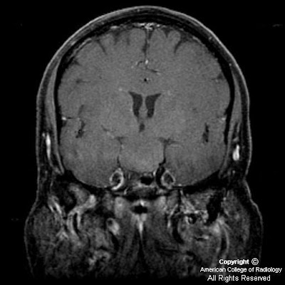
Figure 3: T1 post-gadolinium coronal image shows a homogeneous mass that does not enhance


Diagnosis: Pallister-Hall syndrome


Pallister Hall syndrome is an extremely rare autosomal dominant disorder first described by Judith Hall and Phillip Pallister, both pediatricians and geneticists, in 1980. The underlying mutation causing this syndrome involves the GLI3 protein which participates in gene expression and early development. The manifestations of this disorder result from both the congenital anomalies associated with the genetic mutation and from the hypothalamic hamartomas. Described congenital anomalies include but are not limited to polydactyly, imperforate anus, bifid epiglottis, and renal abnormalities. Despite variability in presentations patients consistently present with polydactyly and frequently with imperforate anus, both of which were included in this patient's medical history.

Hypothalamic hamartomas typically involve a very specific region of the hypothalamus called the tuber cinereum, which consist of gray matter situated between the mammillary bodies and optic chiasm. This region of the brain secretes histamine in association with circadian rhythms. It is most easily identified on sagittal T1 sequences. MRI remains the best imaging modality for appreciating the position and characteristics of hamartomas. The classic image findings are a homogeneous mass in the region of the tuber cinereum which is isointense to gray matter and does not enhance after the administration of gadolinium. Following gadolinium administration, normal pituitary tissue should enhance and be easily identified separate from the tumor. Coronal sequences best demonstrate the mass extending from the floor of the third ventricle. On CT a mass which is isodense to gray matter may be seen in the suprasellar cistern, depending on the size of the lesion. The lesions typically do not calcify.

While specific treatment does not exist for Pallister-Hall syndrome certain measures should be taken in these patients. First, due to the autosomal dominant nature of the disease genetic counseling should be provided to these patients especially if considering conception. Considering the benign nature of the hypothalamic hamartomas, regardless of any syndrome association removal should be based on severity of symptoms. Patients who elect to not have the tumor removed should be evaluated regularly for visual disturbances, neurological changes, and hormonal derangement. Furthermore, consideration should be made for routinely scanning any patient with polydactyly for hypothalamic hamartomas, especially if the patient has other congenital anomalies.