Showing posts with label Oral Surgery. Show all posts

Wednesday, December 14, 2011

Notes on Sialolithiasis-Clinical features, Investigations, Histopathology and Treatment


Sialolithiasis
Thereis presence of one or more round or oval calcified structures in the duct ofthe major or minor salivary glands( salivary stones)

Howthe stone is formed:
Itis assumed that mucin proteins and desquamated ductal epithelial cells form asmall nidus on which the calcium salts are precipitated, this nidus then allowsconcentric lamellar crystallizations to occur and thus sialolith increases insize as a layer by layer gets deposited on it

Clinicalfeatures of sialolithiasis:
About80%of sialolith affects the major salivary glands and there is more predilectionsfor the submandibular gland.
Stonesare rare in children the average age is the 4th decade with no sex preference.
Theyare asymptomatic discovered on dental radiographs.
Ifsymptomatic the chief complains are pain and swelling . Swelling is results asthere is ductal dilatation caused by the ductal blockade.
Thepain is described as pulling drawing or stinging.


SialolithiasisInvestigations:
Panoramicradiograph.
Ultrasound imaging
orsailography

Histopathologyof sialolithiasis:
Stone: On gross examination moststones are yellow or white in colour. they may be round to oval
  - some of the stones are nodular
  - after decalcification the stone showsconcentric rings as of the annual rings of a tree trunk
   -The stone is acellular and amorphous innature and may contain microbial colonies.
Ducts: the ductal lining thatsurrounds sialolith shows variety of reactive changes.
   - there is squamous and mucus cell
     metaplasia and changes to stratifiedsquamous epithelium with numerous mucous goblet cells


Sialolithiasistreatment:
  • Manyof the major salivary gland sialoliths can be removed by manipulation of thestone through major duct orifice
  • Whenmanipulation fails then a surgical cut is made into the main duct
  • Intriangular, or multiple stones and long standing obstructions removal of thestone and sialadenectomy is done.

Saturday, December 10, 2011

Notes on Mucocele and Mucous Retention cyst-Etiology,Clinical Features,Differential Diagnosis and Treatment


Salivary glands react to injury or obstruction by undergoing atrophic degeneration and necrosis with replacement of the parenchyma byinflammatory cells and ultimately fibrous scar formation

Mucocele
It is a tissue swelling composed of pooled mucus thatescapes into the connective tissue from several excretory ducts

Mucocele
Whensalivary duct is severed the acinar cells will continue to secrete saliva intothe severed duct.
Atthe site of the cut/severance the secretory product escape into the connectivetissue forming a pool of mucus that distends the surrounding tissue.


Etiology:
Minorglands of the lip are most prone to severance as a result of injury or bitingthe mucosa.
Intraoral minor salivary can also be effected as result of some irritation as well.

Clinicalfeatures
Mostlyencountered in children and young adults.
Twothird of the mucoceles occur in the 3rd decade of life.
Bothmales and females are effected equally.
Site:mucosal surface of the lower lip
              buccal mucosa
              floor the mouth
              ventral of the tongue and palate

Clinicalappearance of the mococele depends on its location within the submucosa
Moresuperficial zones of mucous extravasations presents a fluctuant mass withbluish translucent appearance.
Patientusually feels the mucocele and the fluctuation in its size
Painis quite rare .
Initiallythe mucocels are well circumscribed but with repeated truma they become nodular,more diffuse and firm on palpation.
Themucoceles have finely vascularized and distended, appearance often referred toas frogs belly that’s why they are also called Ranulas
Whenpart of this ranula is deep seated in to the sumental or submandibular spacethen the term used is the” Plunging Ranula”


Differentialdiagnosis
  • Mucoepidermoidcarcinoma
  • Cavernoushemangeoma (when there is hemorrhage)
  • Blistersseen in some bullous and desqumative disease.
Histopathology:

Underlyingpool of mucin distends the sarface epithelium.
Themucin is walled of by the rim of granulation tissue or in long standing casesby condensed collagen.
Anepithelial lining is lacking
Themucinous material  basophilic oracidophillic and contains  neutrophilsand large oval foam cells the histocytes .
Thebase of the mucocele will reveal feeder duct.
Longstanding mucoceles will show acinar degeneration with fibrosis and minimalinflammation .


Treatment:
Minorsalivary gland mucocele will not resolve on its own it must be surgicallyexcised.
Tominimize the chances of recurrence the feeder gland should also be removed.
Postsurgical parasthesia might occur when the branches of the mental nerve aresevered

Surgical Removal of Mucocele-Video


Mucusretention cyst
Itis a swelling caused by an obstruction of a salivary gland excretory ductresulting in an epithelial lining cavity containing mucus. Mucus retention cystis sometimes also referred as Sialocyst
Themucus retention cyst is lined by epithelium and rarely occur in the majorsalivary gland, when they do occur they are multiple i.e. poly cystic diseaseof the parotid gland


Clinicalfeatures:
Encounteredin adults from 3rd -5th decade.
Thelesion is painless and fluctuant and at times bluish in appearance.
Site:parotid cysts are located in the   superficial lobe as fluctuant well defined mass.
    -with in the oral cavity the floor of themouth is the most common place.
    -this is followed by the lip and the buccalmucosa

Histopathology:
Theepithelium of the cyst is stratified cuboidal or columnar duct like epithelium.
Thecytoplasm in the of these cells is either clear or eosinophlic and my show somefeatures mucous differentiation
70%of these cyst are unilocular rest of the 30% have multilocular pattern.
 
Treatment:
Simpleexcision is the treatment of choice with caution of rupturing the cystic sacs.
Recurrenceis rare.
Howeverdamage to the adjacent gland may result in a mucocele formation.

Friday, December 2, 2011

Notes on Developmental defects of the oral and maxillofacial regions

Developmental defects of the jaws
  1. Orofacial clefts
  2. Coronoid hyperplasia
  3. Condylar hyperplasia
  4. Condylar hypoplasia
  5. Bifid condyle
  6. Exostoses
  7. Torus palatinus
  8. Torus mandibularis
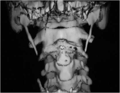
  9. Eagle syndrome
  10. Stafne defect
  11. Hemihyperplasia
  12. Progressive hemifacial atrophy
  13. Segmental odontomaxillary dysplasia
  14. Crouzon syndrome
  15. Apert syndrome
  16. Mandibulofacial dysostosis
  17. Pierre Robin syndrome
Orofacial clefts
  • Most common major congenital defects in humans
  • Frequency of CL +/- CP : Native americans,Asians, Whites,Blacks
  • CL+CP 45%, CPO 30%, CLO 25%
  • More common in males than in females
  • CL: 80%-unilateral (70%-left side), 20%-bilateral
  • 70% of unilateral CLs are associated with CP
  • Minimal manifestation of CP is a bifid uvula
  • Submucosal palatal-the surface mucosa is intact, but adefect exists in the underlying musculature of the soft palate
 
Coronoid hyperplasia
  • Rare, unknown cause
  • Male-to-female ratio = 5:1
  • Endocrine influence, heredity
  • Bilateral CH is more common than unilateral CH*
  • Restricts mandibular opening and causes deviation towardaffected side*
Condylar hyperplasia
  • Excessive growth of one of the condyles
  • Endocrine disturbances and trauma
  • Facial asymmetry, prognathism, crossbite and open bite
  • Most commonly found in adolescents and young adults
  • Self limiting condition


Condylar hypoplasia
  • Underdevelopment of the mandibular condyle
  • Congenital
  •    (mandibulofacialdysostosis, hemifacial microstomia)
  • Acquired
  •   (trauma to thecondylar region during infancy or childhood, infection, radiation therapy, RA,OA)
Bifid condyle
  • Double-headed mandibular condyle
  • Medial and lateral head divided by an anteroposterior groove
  • Trauma, abnormal muscle attachment, teratogenic agents,persistence of a fibrous septum within the condylar cartilage
  • Popping or clicking sound when opening the mouth


Exostoses
  • Localized bony protuberances that arise from the corticalplate
  • Adults
  • A bilateral row of bony hard nodules along the facial aspectof the maxillary and mandibular alveolar ridge


Torus palatinus/mandibularis
Torus palatinus
  • Common exostosis that occurs in the midline of the vault ofthe hard palate
  • Genetics vs environmental or both
  • Asian and Inuit
  • Female:Male = 2:1


Torus mandibularis
  • A bony protuberance along the lingual aspect of the mandibleabove the mylohyoid line in the region of the premolars
  • Bilateral
  • May appear on periapicalradiographs as a radiopacity superimposed on the roots of the tooth


Eagle syndrome
  • Elongation of the styloid process or mineralization of thestylohyoid ligament complex
  • Adults, Most commonly unilateral
  • Vague, radiated facial pain while swallowing, turning thehead or opening the mouth
  • Classic Eagle syndrome – After tonsillectomy
  • Stylohyoid syndrome – impinge on the internal or externalcarotid arteries and associated sympathetic nerve fibers


Stafne defect
  • An asymptomatic radiolucency below the mandibular canal inthe posterior mandible, between the molar teeth and the angle of the mandible
  • Typically well-circumscribed with a sclerotic border
  • Mostly unilateral
  • Reported in middle-aged and older adults, children is rarelyaffected
  • Normal submandibular gland tissue
  • No treatment required


Hemihyperplasia
  • Asymmetric overgrowth of one or more body parts (right side)
  • 2:1 female-to-male predilection
  • Asymmetry may be noted at birth
  •  The enlargementbecomes more accentuated with age especially atpuberty
  • Skin on the affected side: thickened, increasedpigmentation, hypertrichosis, telangiectasias or nevus flemmeus
  • Oral: Macroglossia, larger mandibular canal-crowns
  • Differentiated from Proteus syndrome, NF1
 
Progressive hemifacial atrophy
  • Atrophic changes affecting one side of the face
  • ?trophic malfunction of the cervical sympathetic nervoussystem, trauma, hereditary
  • Close relationship with localized scleroderma
  • Starts during the first 2 decades of life
  • Atrophy of the skin and subcut. Affecting the dermatome ofCNV, osseous hypoplasia
  • Oral: mouth deviated toward the affected side, unilateralatrophy of the tongue, unilateral posterior open bite, deficient root formationor resorption


Segmental odontomaxillary dysplasia
  • Childhood
  • Painless, unilateral enlargement of the maxillary bone alongwith fibrous hyperplasia of the overlying gingival soft tissues
  • Missing developing maxillary premolars, hypoplastic primaryteeth
  • X-Ray: thickened trabeculae-relatively radiopaque, granularappearance


Crouzon syndrome
  • Craniosynostosis : premature closing of the cranial sutures
  • FGFR2 gene mutation on chromosome 10q26
  • Brachy-scapho-trigonocephaly
  • Shallow orbit-visual impairment or total blindness
  • Headaches, beaten metal skull
  • Underdeveloped maxilla
Apert syndrome
  • Characterized by craniosynostosis
  • FGFR2 gene on chromosome 10q26
  • Autosomal dominant, paternal origin
  • Tower(clover leaf) skull, ocular proptosis withhypertelorism, visual loss, hypoplastic middle face – mandibular prognathism,open- mouth appearance, syndactyly*, mental retardation, cleft soft palate


Mandibulofacial dysostosis
  • Defects of structures derived from the first and secondbranchial arches
  • Autosomal dominant
  • TCOF1 gene mapped to chromosome 5q32-q33.1
  • Hypoplastic zygoma, narrow face, depressed cheeks, coloboma, tongue-shaped sideburns, anomalies ofears, underdeveloped mandible

Pierre Robin syndrome
  • Triad: CP, mandibular micrognathia and glossoptosis
  • The retruded mandible results in: Posteriordisplacement of the tongue
                                Lackof support of the tongue    musculature
                                Airwayobstruction
*             Respiratorydifficulty in supine position noted from birth
*             Thepalatal cleft is often U0shaped and wider than isolated CP


Wednesday, November 23, 2011

The Temporomandibular System and Temporomandibular Disorders

Temporomandibular disorder or TMD is a broad term referring to problemswith the jaws and their functioning. It is commonly referred to as"TMJ".This system is very complex and problems are caused by avariety of factors. This web page will attempt to give you a general overviewof TMD so you can better understand the problems you may have been having.

The TemporomandibularSystem

The temporomandibularsystem consists of three basic components; the temporomandibular joint or TMJ,the teeth, and the neuromuscular system. 
Muscles of Mastication and TMJ

TMJ- This is the term often used to describe TMD, but itspecifically refers to the joints that work your lower jaw, or mandible. Thesetwo joints are found just in front of the ears. Their close proximity to theear is why some patients experience ear symptoms and can often hear the jointpop or crack. These two joints never act alone, that is, when your jawfunctions, both joints will be working. There is a small cartilage disc betweenyour lower jaw and skull in the joint. Some TMD problems are caused bydysfunction of this disk. When healthy it acts as a sort of "shockabsorber" for the joint.

Teeth- The teeth are like the third leg of a tripod, theTMJ’s being the other two legs. The alignment of your bite and the functioningof the TMJ’s are intimately connected. Problems in any of the three areas mayaffect the other two.

Neuromuscular- This system is the nerves and muscles whichwork the temporomandibular system. It is important to realize that only thelower jaw, or mandible, moves during jaw function. The upper jaw, or maxilla,does not move; it is firmly attached to the skull. Therefore, the nerves andmuscles are involved in moving the lower jaw only. The nerves transmit themessages for the muscles to move the jaw. They also transmit pain signals tothe brain causing you to feel discomfort. The muscles are fairly large and arefound from the side of your head down to your neck. Other related muscles thatdo not specifically work the jaw, but are sometimes symptomatic are found inthe neck, face, and upper back.

What Defines TMD 
A wide spectrum of specific and non specific disorders thatproduce symptoms of pain and dysfunction of muscles ofmastication,Temporomandibular joint and other associated structures.

Symptoms andsigns of TMD can include some or all of the following.
Jaw pain and/or stiffness
Headaches, usually at the temples and side of head
Vague tooth soreness or toothaches which often move around the mouth
Sensitive teeth
Painful or tender jaw joint
Difficulty opening jaw
Pain and fatigue when eating hard or chewy foods
Clicks, pops, or grinding sound in jaw joint
Ear pain
Cervical neck tension and pain
Tooth wear

Diagnostic classification of Temporomandibular Disorders

Congenital or Developmental Disorders
  • Aplasia 
  • Hypoplasia 
  • Hyperplasia 
  • Neoplasia
Disc Dearrangement Disorders
  • Disc displacement with reduction 
  • Disc displacement without reduction
TMJ dislocation

Inflammatory disorders
  • Capsulitis/Synovitis 
  • Polyarthritides
Osteoarthritis
  • Primary osteoarthritis 
  • Secondary osteoarthritis
Masticatory Muscle disorders
  • Myofacial Pain 
  • Myositis 
  • Myospasm 
  • Myofibrotic contracture 
  • Neoplasia
Temporomandibular disorders come in many forms and varyingdegrees of severity. Basically TMD is a problem when you either experience painand/or a loss of jaw function. The pain can range from a mild ache in themorning to a chronic debilitating pain. Loss of function can be mild jawstiffness to being unable to open the jaw barely at all.
The symptoms of TMD can be an obvious jaw pain in themuscles near the mouth, but can also often exhibit as headache at the temples("temporal headache"). This is due to inflammation in the large jawclosing temporalis muscle which is on either side of your head. To find thismuscle place your hands on your temples and clench your teeth hard and you willfeel this powerful muscle contract. Patients often feel this symptom is atension headache or sinus headache but it can be easily differentiated during aTMD exam. Patients understandably (and many doctors) fail to make thisconnection between TMD and headaches.

The pain of TMD can come from either the muscles or the TMjoint itself; often it comes from a combination of the two. The muscles canache due to causes discussed below. Pain in the joint is usually due toinflammation within the structure itself. Sometimes the symptoms are found inother facial structures; such as dull ear pain, toothache, neck pain, etc. Thisreferred pain is fairly common but it is important to rule out medical and/ordental causes of these pains prior to TMD treatment.
Loss of jaw function can be due to muscular and/or jointproblems as well, just as pain can. The degree to which each is involved isdetermined through thorough history and exam.

Causes of TMD
Temporomandibular disorders rarely have a single cause. Anyone of the following factors may contribute to TMD. Each patient presents withan individual combination of factors that are determined during history takingand clinical exam.

Trauma - Acute trauma to the jaws such as a car accident, afall, a punch, etc. can cause damage to the muscles and/or joint. The acutepain and loss of function is usually responsive to conservative treatment.Sometimes trauma to the joint can cause chronic damage which may eventuallycontribute to a TMD problem at a later time.

Bruxism - Bruxism refers to a non functional grinding andclenching of the teeth. Some do this while awake but more often it is donewhile sleeping. Most people grind their teeth while sleeping to some degree.For whatever reason some people do this very hard to the point where they wearthe enamel from their teeth. This bruxing is done by the jaw muscles and by themorning they can be painful due to fatigue. This constant pressure also candamage the TMJs over time. Bruxism is the most common factor found in TMD.

Malocclusion - This term means "bad bite".Sometimes when the teeth do not bite together in harmony with the shape and positionof the joints it can place pressure on the joints. Missing teeth can sometimescontribute to this as well. The misalignment can also put strain on the jawmuscles. This factor can be mild to severe. Though the bite is an importantpart of the whole system it is only altered after conservative measures andonly if it is felt improvement will result. In some patients discrepancies(known as interferences) become apparent after wearing the NTI device for a fewmonths and a bite adjustment is recommended. Treatment of the bite for TMD isusually not needed but its contribution to the whole must be examined.

Emotion - Emotional stress often plays an integral role inthe development of TMD. This occurs due to two basic reasons. Stress increasesboth the severity and duration of bruxism while asleep. Also, many willsubconsciously clench and/or grind their teeth more while awake during times ofstress. The other way stress contributes to TMD is that during times of stressyour adaptability and pain threshold will go down. As a result you are morelikely to experience symptoms of TMD if other factors already exist (bruxing,joint damage, etc.).
Emotional conditions beyond daily life stress can contributeto TMD as well. Depression, anxiety disorders, and the like can often have TMDproblems arise. These conditions are quite stressful and it is not hard toimagine why TMD would develop.

Ergonomic - Your job and how you do it can contribute to TMDand related problems. If you work at a computer all day, for example, you maybe holding your head in such a way that places strain on your TM system.

TMD Treatment
Treatmentplans for TMD are as varied as the patients that present with it. Each patientmust be treated differently depending on the uniqueness of their problems andthe contributing factors. Pain Relief Info.
It is veryimportant to realize that the goal of TMD treatment is to minimize pain andestablish a return to function. TMD conditions are not "cured" butare managed instead. The basic goal is to allow the muscles and joints to healthrough rest and care. Often damage to the joint itself can not be reversed,but the body can often heal it enough to return to function without pain. Wealso want to teach you to recognize the symptoms early and manage them yourselfonce we give you the tools to do so. This condition can often recur later onbut early care can minimize the severity.
The basicphilosophy of treatment is to do the conservative and reversible treatmentsfirst. Irreversible treatments, such as surgery or orthodontics, are onlyconsidered if conservative steps have failed to bring lasting relief. Thesemore radical treatments are rarely used. Most patients respond well to simplercare.

Thefollowing treatment modalities may be used in each case.

Conservativetreatment
  • Patienteducation and self care 
  • Rest andrelaxation 
  • Cognitive behavioralintervention 
  • Pharmacotherapy 
  • Occlusaltherapy 
  • Occlusal therapy 
  • Orthopedic appliances 
  • Physiotherapy 
  • Rehabilitationof Denatal deficits 
  • Managementof trigger points

SurgicalTreatment
  • Arthrocentesis 
  • Arthroscopy 
  • Arthroplasty 
  • Discectomy 
  • Condylectomy


Occlusal Splint - Also called a night guard is designed toprotect the teeth from further wear. These also will reduce the severity ofgrinding at night and allow the muscles to rest. In more severe cases it needsto be worn all day as well to allow the TMJ and muscles to rest.

Wednesday, November 9, 2011

Anatomy and Physiology of the Salivary Glands and Sialography


Types of salivaryglands

The Major Salivary Glands
  • Parotid
  • Submandibular
  • Sublingual
The Minor SalivaryGlands
 
Embryology
  • 6th-8th Weeks ofGestation
  • Parotid
    • First to develop
    • Last to becomeencapsulated
  • Autonomic NervousSystem Crucial
Anatomy of ParotidGland
  • Wedge shaped with 5processes
    • 3 Superficial
    • 2 Deep
  • Parotid Compartment
    • Superior – Zygoma
    • Posterior – EAC
    • Inferior – Styloid,ICA, Jugular Veins
  • 80% overlies
  • Masseter &Mandible
  • 20% Retromandibular
  • Stylomandibular,Tunnel,Isthmus of Parotid
  • Tail of Parotid
Parapharyngeal Space
  • Prestyloid Compartment
  • PoststyloidCompartment (Paragangliomas)
Stensen’s Duct
  • Arises from anteriorborder
  • 1.5 cm inferior toZygomatic arch
  • Pierces Buccinator at2nd Molar
  • 4-6 cm in length
  • 5 mm in diameter
Parotid Capsule
  • Superficial layer DeepCervical Fascia
  • Superficial layer
  • Deep layer


CN VII-Facial nerve
  • 2 Surgical zones
  • 3 Motor branches
  • immediately
  • Pes Anserinus – 1.3 cm
  • Temperofacial Division
  • Cervicofacial Division
  • 5 Terminal branches
Localization of CN VII
  • Tragal pointer
  • Tympanomastoid suture
  • Posterior bellyDigastric
  • Styloid process
  • Retrograde dissection
  • Mastoidectomy
  • Great Auricular nerve
  • Auriculotemporal nerve
    • Superficial Temporalvessels
    • Frey’s Syndrome
Neural compartment-VII, Great Auricular,Auriculotemporal
Venous compartment-Retromandibular vein
Arterial compartment-SuperficialTemporal/Transverse Facial

Lymphatics
  • Paraparotid &Intraparotid nodes
  • Superficial & DeepCervical nodes
Submandibular Gland-Anatomy
 
  • The ‘Submaxilla’
  • Submandibular Triangle
  • Mylohyoid ‘C’
  • Marginal Mandibular branch
  • Capsule fromsuperficial layer of Deep Cervical fascia
Wharton’s duct
  • Exits medial surface
  • Between Mylohyoid &Hyoglossus
  • 5 cm in length
  • Lingual nerve & CNXII
Innervation
Superior CervicalGanglion (symp)
Submandibular Ganglion(para)
Artery: Submentalbranch of Facial a.
Vein: Anterior Facial Vnerve.
Lymphatics: DeepCervical and Jugular chains
Facial artery nodes
Between Mandible &Genioglossus
No capsule
Ducts of Rivinus +/-Bartholin’s duct
Sialogram not possible
Innervation: Same asSubmandibular
Artery/Vein: Sublingualbranch of Lingual & Submental branch of Facial
Lymphatics:Submandibular nodes

Minor Salivary Glands

  • 600-1,000
  • Simple ducts
  • Buccal, Labial,
  • Palatal, Lingual
  • Tumor sites:
  • Palate, upper lip,cheek
  • Lingual & Palatinenn.
Imaging of SalivaryGland-Important things to remember
  • CT – Inflammatory
  • MR – Tumor
  • Children: U/S & MR
  • NO sialogram duringactive infection
  • Parotid is fatty
The Secretory Unit
  • Acinus (serous, mucous,mixed)
  • Myoepithelial cells
  • Intercalated duct
  • Striated duct
  • Excretory duct
Microanatomy of Salivary glands
  • Striated &Intercalated ducts well developed in serous, NOT mucous glands
  • Striated duct: HCO3into, Cl from lumen
  • Intercalated duct: Kinto lumen, Na from lumen, producing hypotonic fluid
  • Excretory ducts do NOTmodify saliva
 
The Bicellular Theory
  • Intercalated duct
  • Excretory duct
The Multicellular Theory

Parotid: serous &fatty
Submandibular: mixed serous
Sublingual: mixed mucous
Stroma: Plasma cells

Function of Saliva
  1. Moistens oral mucosa
  2. Moistens & cools food
  3. Medium for dissolvedfood
  4. Buffer (HCO3)
  5. Digestion (Amylase,Lipase)
  6. Antibacterial (Lysozyme, IgA, Peroxidase, FLOW)
  7. Mineralization
  8. Protective Pellicle
 
Effects of Salivaryhypofunction
  • Candidiasis
  • Lichen Planus
  • Burning Mouth
  • Aphthous ulcers
  • Dental caries
  • Xerostomia not reliable
 
Production of Saliva

  • Primary secretion
  • Ductal secretion
  • The “secretory potential”
  • (hyperpolarizes)
  • Increased flow rate yieldsdecreased
  • hypotonicity & K
Autonomic Innervation
Parasympathetic
  • Abundant, waterysaliva
  • Amylase down
Sympathetic
  • Scant, viscous saliva
  • Amylase up
Salivary Flow
  • 1-1.5 L/day (1 cc/min)
  • Unstimulated state
  • Submandibular
  • Stimulated state
  • Parotid
  • Sublingual & minor
  • Mucin
Effects of Aging
Total salivary flowindependent of age
Acinar cellsdegenerate with age
Submandibular glandmore sensitive to metabolic/physiologic change
Unstimulated salivaryflow more greatly affected by physiologic changes

Sialography
Radiologic examinationof the salivary glands
The submandibular andparotid glands are investigated by this method
The sublingual glandis usually not evaluated this way-Difficulty incannulation

Indications
  • Ductal obstruction-Stonesor tumors
  • Inflammation of a ductor gland
Contraindications
  • Severe infection of agland
  • Known allergies tocontrast media
Equipment
  • Fluoroscopic unitw/spot film capabilities
  • Cannula forintroducing contrast
  • Connecting tubing
  • Lemons
  • Dilators for duct
  • 5 mL syringe
  • Overhead light
  • Gauze
  • Contrast
Preliminary andProcedure Radiographs
  • Parotid-Tangential
    • Perpendicular tocassette, directed to lateral surface of mandibular ramus
  • Submandibular-Lateral
    • Perpendicular tocassette, directed to 1 in. superior to mandibular angle to demonstrate parotidgland
    • Inferior margin ofmandibular angle to demonstrate the submandibular gland
Patient Preparation
  1. Thorough explanationof examination
  2. Any removable dentalwork, jewelry, and other artifact causing opaque items must be removed
  3. Consent must be signed
Procedure
  • The patient firstsucks on a lemon wedge to open the ducts
  • An overhead lamp isused to provide adequate light
  • The duct iscannulated, not punctured, and contrast is introduced with fluoroscopicguidance
  • Radiographs are obtained
  • After the radiographs,the patient then sucks on a lemon wedge to evacuate the contrast
  • Obtain post-procedureradiographs as indicated
Lateral Parotid GlandRadiograph


Lateral SubmandibularGlands