Showing posts with label Systemic. Show all posts

Monday, March 7, 2011

Chloroma of epidural space


Additional clinical history: Acute leukemia.


Findings

Large anterior epidural mass extending from posterior clinoid to the cervicothoracic junction measuring approx. 6x3x1.5 cm with mass effect on the anterior pons, medulla, and upper cervical cord. No post-contrast imaging obtained.

Differential diagnosis: Epidural mass
- Metastatic disease
- Lymphoma
- Leukemia/chloroma
- Chordoma
- Osteomyelitis/epidural abscess
- Epidural hematoma
- Primary tumor such as neurofibroma/schwannoma


Diagnosis: Chloroma of epidural space


Key points

AKA granulocytic sarcoma, extramedullary myeloblastoma.
Most commonly occurs in the setting of AML.
Can also occur in setting of chronic myelogenous leukemia and other myeloproliferative disorders.
These tumors can involve any part of the body, either concurrently or sequentially.

Imaging characteristics:
- NECT: Isodense or hyper dense to brain or muscle
- MR: Hypo intense or Iso intense on T1-weighted MR images, heterogeneously Iso intense or hyper intense on T2-weighted MR images
- MR+C: Enhance homogeneously after injection of contrast medium

Paraspinal and intraspinal lesions are also thought to arise from perivenous arachnoid spread of leukemic cells. Uncommonly, spinal involvement by granulocytic sarcoma may cause compression of the spinal cord, cauda equina, or nerve roots

Tuesday, December 7, 2010

Methotrexate neurotoxicity


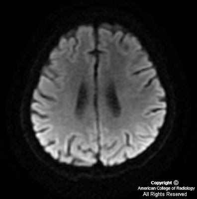
Additional clinical history: Patient was diagnosed with acute lymphocytic leukemia 2 months previously. He is status post induction therapy with a negative bone marrow biopsy, and is currently receiving consolidation chemotherapy with methotrexate, and presents with right upper extremity weakness.


Findings

MR images of the brain demonstrate a focal area of diffusion restriction involving the left frontoparietal white matter. There is minimal associated T2/FLAIR hyperintensity. No associated enhancement. Remainder of the brain was within normal limits.
Imaging done four months later shows lesion has nearly resolved.


Diagnosis: Methotrexate neurotoxicity


Discussion

Methotrexate is a folic acid analogue. Its cytotoxic effects are carried out through inhibition of the enzyme dihydrofolate reductase, which reduces tetrahydrofolic acid levels, ultimately inhibiting cell division.

From bone marrow cell precursors to the quickly dividing cells of the intestinal tract, methotrexate exerts its effects on all dividing cells in the body. One of its rare side effects is CNS toxicity. The decreased folate levels achieved with methotrexate have implications on metabolism of adenosine, homocysteine, and biopterin. Low folate levels lead to a subsequent decrease in S-adenosyl-methionine(SAM) concentrations. This eventually leads to chronic demyelination and neurologic symptoms.

An additional side effect of MTX is the elevated levels of adenosine in the CSF. Adenosine is a vasodilator, which causes dilatation of cerebral vasculature resulting in neurotoxicity. The increased homocysteine levels caused by MTX have been shown to damage vascular endothelium and lead to subsequent strokes and thromboemboli. Methotrexate has also been found to cause cytotoxic edema, which is the most common cause of lesions that enhance on MRI DWI.

The neurotoxicity caused by MTX can be immediate, acute to subacute, or delayed. Symptoms of the disease can range from headache, nausea, vomiting, and fever, to transient or permanent focal neurologic symptoms. The immediate form occurs within a day of MTX administration and presents as a chemical meningitis. The acute to subacute form presents from days to weeks after administration of MTX, and presents with seizures or focal neurologic symptoms. The delayed form presents as leukoencephalopathy and a generalized decrease in higher cognitive function.


Radiological findings

A case series containing nine cases of MTX neurotoxicity revealed that lesions found in this disease tend to be focal and show up on DWI as well as T2 and FLAIR imaging. These abnormalities can continue to persist on imaging long after the symptoms have resolved. The DWI shows diffusion restriction with T2/FLAIR hyperintensity being less conspicuous.

In another independent case study on MTX neurotoxicity, MRI demonstrated restriction diffusion with no significant T2 or FLAIR signal abnormality. Based on a combination of these imaging findings, it was determined that cytotoxic edema was likely the cause of focal neurologic symptoms on the patient, and demyelination was a less likely cause based on the MRI findings.

A different case study had MRI findings showing subtle signal changes in the left centrum semiovale, with an obviously abnormal area of restricted diffusion, indicating the presence of increased fluid. The authors of this case also mentioned a relation between elevated choline levels in lesion areas with myelin breakdown.

The lesion in this disease is similar in appearance to ischemic stroke, but differs in distribution. The lesions in MTX neurotoxicity can show up in many different patterns, whereas ischemic strokes often follow a vascular distribution, helping differentiate the two.


Radiology

MRI:
MRI with DWI is the gold standard for diagnosis
Will show focal areas of demyelination and/or edema throughout the brain
Can be normal, even in the presence of symptoms
Must perform early to avoid unnecessary workup


CT:
Can be used to rule out other etiologies that may cause focal symptoms, but is not a sensitive test for demyelination and edema found with MTX neurotoxicity
Ultimately need MRI to make diagnosis as CT is often negative
Angiography
Not very useful as it is usually normal

Wednesday, November 10, 2010

Methotrexate (MTX) induced transient neurotoxicity

14-year-old child with history of Acute Lymphoblastic Leukemia (ALL), on induction chemotherapy complaining of left sided weakness and facial asymmetry of acute onset.


The patient was given supportive treatment and aminophylline, symptoms resolved and an MRI was repeated after 3 days.


Findings

Diffusion weighted images with corresponding ADC maps show restricted diffusion involving bilateral centrum semiovale (Figure 1 and Figure 2).
Diffusion weighted images with corresponding ADC maps, from the MRI done after clinical improvement, show resolution of abnormalities seen on DW and ADC Maps in the initial study (Figure 3 and Figure 4).


Diagnosis: Methotrexate (MTX) induced transient neurotoxicity


With improvements in antileukemic treatment there has been a steady increase in long term survivors of ALL. However a myriad of neurological complications are seen during and after treatment. These maybe broadly categorized into those related to chemotherapeutic agents, radiation therapy, coagulopathy, immunosuppression and marrow transplantation.

Methotrexate is an essential component of treatment regimens in ALL. It can be administered both intravenously and intrathecally. Though hematologic and mucocutaneous consequences are more common, the CNS adverse effects are more worrisome. Chronic leukoencephalopathy as a result of methotrexate and radiotherapy is a well recognized complication and usuallly associated with cognitive deficits rather than focal neurologic deficits although subacuteacute encephalopathy after methotrexate may occur as well and usually presents as headache, confusion, disorientation, seizure, and focal neurologic deficit. A vast majority of patients show hemiparesis and aphasia.

High level of adenosine is thought to be responsible for methotrexate induced toxicity. Statistically the periventricular white matter is the most common area affected. On diffusion weighted imaging these areas show increased signal intensity and hypointensity on corresponding apparent diffusion coefficient (ADC) maps. There may be no abnormality identified on T1, T2, and FLAIR sequences during the acute symptomatic phase.

Clinical resolution is followed by the appearance of residual FLAIR hyperintensities in the involved areas, which show gradual regression. There is no established treatment, however several anecdotes report symptomatic resolution with aminophylline therapy.

Monday, November 8, 2010

Neuroblastoma metastases


Findings

Figure 1: Axial post gadolinium T1 weighted image showing solid enhancing parenchymal lesion in the right temporal lobe with dural and leptomeningeal disease.
Figure 2: Coronal post gadolinium image showing dural enhancement along the tentorium and leptomeningeal enhancement.
Figure 3: Axial susceptibility weighted imaging revealing the hemorrhagic nature of the lesion.


Diagnosis: Neuroblastoma metastases


Neuroblastoma metastatic to the central nervous system is extremely rare, and the reported incidence varies from 1% to 16% at recurrence. Paediatric tumors that metastasise to the brain, in order of frequency, include neuroblastoma, osteosarcoma, Ewing sarcoma, rhabdomyosarcoma and Wilm tumor.

Risk factors for developing intracranial metastases include lumbar puncture at diagnosis, ages 2 to 3 years, bone marrow involvement, and MYCN gene amplification. Newer chemotherapeutic agents with better activity fail to penetrate the blood-brain barrier, thus facilitating a sanctuary for tumor cells within the central nervous system. As a result, the metastases evolve and become extensive before becoming clinically evident. Metastatic spread of tumor cells to central nervous system may occur either via hematogenous or cerebrospinal fluid routes and involve the neuroparenchyma, leptomeninges or dura.

Neuroparenchymal metastases from neuroblastoma have varied appearances. They may be cystic lesions with calcified mural nodules. The wall and mural nodules show intense enhancement with contrast. Metastases may also be solid and hemorrhagic and show homogeneous enhancement. Gradient or susceptibility weighted imaging would help in detecting hemorrhagic components. Leptomeningeal and dural metastatic involvement if present indicates poor prognosis.