Epidermolysis Bullosa
Etiology
• A diverse group ofpredominantly cutaneous, but also mucosal, mechanobullous diseases
• Inherited form:autosomal dominant or recessive patterns may occur
• Acquired form(acquisita): autoimmune from autoantibodies (immunoglobulin G [IgG]) to typeVII collagen deposited within the basement membrane zone and upper dermis orlamina propria
Clinical Presentation
• Variable, dependingupon the specific form of many subtypes recognized
• Mucosal lesionsrange in severity from mild to debilitating, depending on subtype:
• Inherited forms havewide range of oral mucosal involvement, with most severe form (autosomalrecessive, dermolytic) also demonstrating enamel hypoplasia and caries
• Acquisita form withmucous membrane pemphigoid variant shows oral and conjunctivalerosions/blisters
• Mucosal involvementabsent in several variants
• Scarring andstricture formation common in severe recessive forms
• Mucosa is oftenfriable, but it may be severely blistered, eroded, or ulcerated.
• Loss of oralanatomic landmarks may follow severe scarring (eg, tongue mucosa may becomesmooth and atrophic with episodes of blistering and scarring).
• Obliteration ofvestibules, reduction of oral opening, ankyloglossia
• Scarring can beassociated with atrophy and leukoplakia, with increased risk for squamous cellcarcinoma development.
Microscopic Findings
• Bullae vary inlocation depending upon the form that is present:
• Intraepithelial innonscarring forms
• Atepithelial–connective tissue junction in dystrophic forms
•Subepithelial/intradermal in scarring forms
• Ultrastructuralfindings are as follows:
• Intraepithelialforms associated with defective cytokeratin groups
• Junctional formsassociated with defective anchoring filaments at hemidesmosomal sites(epithelial–connective tissue junction)
• Dermal typesdemonstrate anchoring fibril or collagen destruction.
Diagnosis
• Distribution oflesions
• Family history
• Microscopicevaluation
• Ultrastructuralevaluation
• Immunohistochemicalevaluation of basement membrane zone using specific labeled antibodies asmarkers for site of blister formation
Differential Diagnosis
• Varies with specificform
• Generally includesthe following:
• Bullous pemphigoid
• Mucous membrane(cicatricial) pemphigoid
• Erosive lichenplanus
• Dermatitisherpetiformis
• Porphyria cutaneatarda
• Erythema multiforme
• Bullous impetigo
• Kindler syndrome
• Ritter’s disease
Treatment
• Acquisita form:
• Some recent successwith colchicine and dapsone
• Immunosuppressiveagents including azathioprine, methotrexate, and cyclosporine may be effective
• Acquisita andinherited forms:
• Avoidance of trauma
• Dental preventionstrategies including extra-soft brushes, daily topical fluoride applications,dietary counseling
Prognosis
• Widely variabledepending on subtype
Erythema Multiforme
Etiology
• Many cases precededby infection with herpes simplex; less often with Mycoplasma pneumoniae or other organisms
• May be related todrug consumption, including sulfonamides, other antibiotics, analgesics, phenolphthalein-containinglaxatives, barbiturates
• Another trigger maybe radiation therapy.
• Essentially animmunologically mediated reactive process, possibly related to circulatingimmune complexes
Clinical Presentation
• Acute onset ofmultiple, painful, shallow ulcers and erosions with irregular margins
• Early mucosallesions are macular, erythematous, and occasionally bullous.
• May affect oral mucosaand skin synchronously or metachronously
• Lips most commonlyaffected with eroded, crusted, and hemorrhagic lesions (serosanguinous exudate)known as Stevens-Johnson syndrome when severe
• Predilection foryoung adults
• As many as one-halfof oral cases have associated erythematous to bullous skin lesions.
• Target or iris skinlesions may be noted over extremities.
• Genital and ocularlesions may occur.
• Usuallyself-limiting; 2- to 4-week course
• Recurrence iscommon.
Diagnosis
• Appearance
• Rapid onset
• Multiple siteinvolvement in one-half of cases
• Biopsy results oftenhelpful, but not always diagnostic
Differential Diagnosis
• Viral infection, inparticular, acute herpetic gingivostomatitis (Note: Erythema multiforme rarelyaffects the gingiva.)
• Pemphigus vulgaris
• Major aphthousulcers
• Erosive lichenplanus
• Mucous membrane(cicatricial) pemphigoid
Treatment
• Mild (minor) form:symptomatic/supportive treatment with adequate hydration, liquid diet, analgesics,topical corticosteroid agents
• Severe (major) form:systemic corticosteroids, parenteral fluid replacement, antipyretics
• If evidence of anantecedent viral infection or trigger exists, systemic antiviral drugs during thedisease or as a prophylactic measure may help.
• See “Therapeutics”section for details.
Prognosis
• Generally excellent
• Recurrences common
Hand-Foot-and-MouthDisease
Etiology
• A very commonenterovirus infection (coxsackievirus A10 or A16), which may occur in mild epidemicproportion, chiefly in children
• Incubation period isshort, usually less than 1 week
Clinical Presentation
• Oral mucosal lesionswith focal herpes simplex–like appearance, usually involving nonkeratinizedtissue (soft palate, floor of mouth, labial-buccal mucosa)
• Accompanying palmar,plantar, and digital lesions are deeply seated, vesicular, and erythematous
• Short course withmild symptoms
Diagnosis
• Concomitant oral andcutaneous lesions
• Skin lesionscommonly involve hands and feet.
• Skin lesions mayinvolve buttocks.
• Antibody-titerincrease measured between acute and recovery phases
Differential Diagnosis
• Herpangina
• Herpes simplexinfection
• Acute lymphonodularpharyngitis
Treatment
• Symptomatictreatment only
• Patient should becautioned against the use of aspirin to manage fever.
Prognosis
• Excellent
• Lifelong immunity,but it is strain specific
Herpangina
Etiology
• Most often bymembers of coxsackievirus group A (7, 9, 10, and 16) or group B (1–5)
• Occasionally due toechovirus 9 or 17
Clinical Presentation
• Incubation period of5 to 9 days
• Acute onset
• Usually endemic inyoung children; usually occurs in summer
• Often subclinical
• Posterior oralcavity, tonsillar pillars involved
• Macular erythematousareas precede short-lived vesicular eruption, followed by superficialulceration
• Accompanied bypharyngitis, dysphagia, fever, malaise, headache, lymphadenitis, and vomiting
• Self-limitingcourse, usually under 2 weeks
Diagnosis
• Other viralillnesses to be ruled out or separated
• Course, time ofyear, location of lesions, contact with known infected individual
Differential Diagnosis
• Hand-foot-and-mouthdisease
• Varicella
• Acute herpeticgingivostomatitis
Treatment
• Soft diet
• Hydration
• Antipyretics
• Chlorhexidine rinses
• Compounded mouthrinses
Prognosis
• Excellent
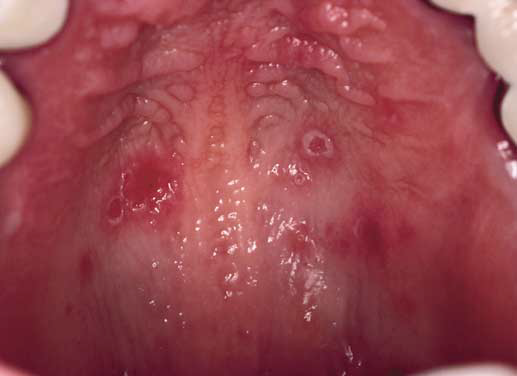
Herpetic Stomatitis:Primary
Etiology
• Herpes simplex virus(HSV)
• Over 95% of oral primary herpes due to HSV-1
• Physical contact ismode of transmission
Clinical Presentation
• 88% of populationexperience subclinical infection or mild transient symptoms
• Most cases occur inthose between 0.5 and 5 years of age.
• Incubation period ofup to 2 weeks
• Abrupt onset inthose with low or absent antibody to HSV-1
• Fever, anorexia,lymphadenopathy, headache, in addition to oral ulcers
• Coalescing, grouped,pinhead-sized vesicles that ulcerate
• Ulcers show ayellow, fibrinous base with an erythematous halo
• Both keratinized andnonkeratinized mucosa affected
• Gingival tissue withedema, intense erythema, pain, and tenderness
• Lips, perioral skinmay be involved
• 7- to 14-day course
Diagnosis
• Usually by clinicalpresentation and pattern of involvement
• Cytology preparationto demonstrate multinucleate virusinfected giant epithelial cells
• Biopsy results ofintact macular area show intraepithelial vesicles or early virus-inducedepithelial (cytopathic) changes
• Viral culture orpolymerase chain reaction (PCR) examination of blister fluid or scraping frombase of erosion
Differential Diagnosis
• Herpangina
• Hand-foot-and-mouthdisease
• Varicella
• Herpes zoster(shingles)
• Erythema multiforme(typically no gingival lesions)
Treatment
• Soft diet and hydration
• Antipyretics (avoidaspirin)
• Chlorhexidine rinses
• Systemic antiviralagents (acyclovir, valacyclovir) if early in course or in immunocompromisedpatients
• Compounded mouthrinse
Prognosis
• Excellent inimmunocompetent host
• Remission/latentphase in nearly all those affected who have adequate antibody titers
Impetigo
Etiology
• Cutaneous bacterialinfection: Streptococcus and Staphylococcus species
• Is spread throughdirect contact
• Highly contagious
Clinical Presentation
• Honey-colored,perioral crusts preceded by vesicles
• Flaccid bullae lesscommon (bullous impetigo)
Diagnosis
• Clinical features
• Culture of organism(usually group A, â-hemolyticstreptococci or group II Staphylococcusaureus)
• Herpes simplex(recurrent)
• Exfoliativecheilitis
• Drug eruptions
• Othervesiculobullous diseases
Treatment
• Topical antibiotics(mupirocin, clindamycin)
• Systemic antibiotics
Prognosis
• Excellent
• Rarely,poststreptococcal glomerulonephritis may develop.
Mucous MembranePemphigoid
Etiology
• Autoimmune; triggerunknown
• Autoantibodiesdirected against basement membrane zone antigens
Clinical Presentation
• Vesicles and bullae(short lived) followed by ulceration
• Multiple intraoralsites (occasionally gingiva only)
• Usually in olderadults
• 2:1 femalepredilection
• Ocular lesions notedin one-third of cases
• Proclivity forscarring in ocular, laryngeal, nasopharyngeal, and oropharyngeal tissues
Microscopic Findings
• Subepithelial cleftformation
• Linear pattern IgGand complement 3 (C3) along basement membrane zone; less commonly IgA
• Directimmunofluorescence examination positive in 80% of cases
• Indirectimmunofluorescence examination usually negative
• Immunoreactantsdeposited in lamina lucida in most patients
Diagnosis
• Biopsy
• Direct immunofluorescentexamination
Differential Diagnosis
• Pemphigus vulgaris
• Erythema multiforme
• Erosive lichen planus
• Lupus erythematosus
• Epidermolysis bullosa acquisita
Treatment
• Topical corticosteroids
• Systemic prednisone,azathioprine, or cyclophosphamide
•Tetracycline/niacinamide
• Dapsone
• See “Therapeutics”section for details.
Prognosis
• Morbidity related tomucosal scarring (oropharyngeal, nasopharyngeal, laryngeal, ocular, genital)
• Management oftendifficult due to variable response to corticosteroids
• Management oftenrequires multiple specialists working in concert (dental, dermatology,ophthalmology, otolaryngology)
Paraneoplastic Pemphigus
Etiology
• Autoimmune,triggered by malignant or benign tumors
• Autoantibodiesdirected against a variety of epidermal antigens including desmogleins 3 and 1,desmoplakins I and II, and other desmosomal antigens, as well as basementmembrane zone antigens
Clinical Presentation
• Short-lived vesiclesand bullae followed by erosion and ulceration; resembles oral pemphigus
• Multiple oral sites
• Severe hemorrhagic,crusted erosive cheilitis
• Painful lesions
• Cutaneous lesionsare polymorphous; may resemble lichen planus, erythema multiforme, or bullouspemphigoid
• Underlying neoplasmssuch as non-Hodgkin’s lymphoma, leukemia, thymoma, spindle cell neoplasms,Waldenström’s macroglobulinemia, and Castleman’s disease
Microscopic Findings
• Suprabasilaracantholysis, keratinocyte necrosis, and vacuolar interface inflammation
• Directimmunofluorescent testing is positive for epithelial cell surface deposition ofIgG and C3 and a lichenoid tissue reaction interface deposition pattern
• Indirectimmunofluorescent testing is positive for epithelial cell surface IgGantibodies
• Special testing withmouse and rat bladder, cardiac muscle, and liver may demonstrate paraneoplasticpemphigus antibodies that bind to simple columnar and transitional epithelia
Diagnosis
• Biopsy of skin ormucosa
• Directimmunofluorescent examination of skin or mucosa
• Indirectimmunofluorescent examination of sera including special substrates
Differential Diagnosis
• Pemphigus vulgaris
• Erythema multiforme
• Stevens-Johnsonsyndrome
• Mucous membrane(cicatricial) pemphigoid
• Erosive oral lichenplanus
Treatment
• Identification ofconcurrent malignancy
• Immunosuppressivetherapy
Prognosis
• Good with excisionof benign neoplasms
• Grave, usuallyfatal, with malignancies
• Management is verychallenging.
Pemphigus Vulgaris
Etiology
• An autoimmunedisease where antibodies are directed toward the desmosome-related proteinsdesmoglein 3 or desmoglein 1
• A drug-induced formexists with less specificity in terms of immunologic features, clinicalpresentation, and histopathology
Clinical Presentation
• Over 50% of casesdevelop oral lesions as the initial manifestation
• Oral lesions developin 70% of cases
• Painful, shallowirregular ulcers with friable adjacent mucosa
• Nonkeratinized sites(buccal, floor, ventral tongue) often are initial sites affected
• Lateral shearingforce on uninvolved skin or mucosa can produce a surface slough or inducevesicle formation (Nikolsky sign)
Microscopic Findings
• Separation orclefting of suprabasal from basal layer of epithelium
• Intact basal layerof surface epithelium
• Vesicle forms atsite of epithelial split
• Nonadherent spinouscells float in blister fluid (Tzanck cells)
• Directimmunofluorescence examination positive in all cases
• IgG localization tointercellular spaces of epithelium
• C3 localization tointercellular spaces in 80% of cases
• IgA localization tointercellular spaces in 30% of cases
• Indirectimmunofluorescence examination positive in 80% of cases
• General correlationwith severity of clinical disease
Diagnosis
• Clinical appearance
• Mucosalmanifestations
• Direct/indirectimmunofluorescent studies
Differential Diagnosis
• Mucous membrane(cicatricial) pemphigoid
• Erythema multiforme
• Erosive lichenplanus
• Drug reaction
• Paraneoplasticpemphigus
Treatment
• Systemicimmunosuppression
• Prednisone, azathioprine,mycophenolate mofetil, cyclophosphamide
• Plasmapheresis plusimmunosuppression
• IVIg for somerecalcitrant cases
• See “Therapeutics”section for details.
Prognosis
• Guarded
• Approximately a 5%mortality rate secondary to long-term systemic corticosteroid–relatedcomplications
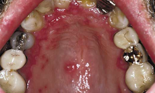
Recurrent HerpeticStomatitis: Secondary
Etiology
• Herpes simplex virus
• Reactivation oflatent virus
Clinical Presentation
• Prodrome oftingling, burning, or pain at site of recurrence
• Multiple, grouped,fragile vesicles that ulcerate and coalesce
• Most common onvermilion border of lips or adjacent skin
• Intraoralrecurrences characteristically on hard palate or attached gingiva (masticatorymucosa)
• In immunocompromisedpatients, lesions may occur in any oral site and are more severe (herpeticgeometric glossitis).
Diagnosis
• Characteristicclinical presentation and history
• Viral culture or PCRexamination of blister fluid or scraping from base of erosion
• Cytologic smear
• Directimmunofluorescence examination of smear
Differential Diagnosis
• Erythema multiforme
• Herpes zoster(shingles)
• Herpangina
• Hand-foot-and-mouthdisease
Treatment
• Acyclovir orvalacyclovir early in prodrome
• Supportive
• Acyclovir may beused for prophylaxis for seropositive transplant patients
• Ganciclovir may beused for human immunodeficiency virus (HIV)-positive patients, especially thoseco-infected with cytomegalovirus.
• For recurrent herpeslabialis, see “Therapeutics” section.
Prognosis
• Excellent
• Healing withoutscarring within 10 to 14 days
• Protracted healingin HIV-positive patients
Stevens-JohnsonSyndrome
Etiology
• A complex mucocutaneousdisease affecting two or more mucosal sites simultaneously
• Most common trigger:antecedent recurrent herpes simplex infection
• Infection with Mycoplasma also may serve as a trigger.
• Medications mayserve as initiators in some cases.
• Sometimes referredto as “erythema multiforme major”
Clinical Presentation
• Labial vermilion andanterior portion of oral cavity usually affected initially
• Early phase ismacular followed by erosion, sloughing, and painful ulceration
• Lip ulcers appearcrusted and hemorrhagic.
• Pseudomembrane;foul-smelling presentation as bacterial colonization supervenes
• Posterior oralcavity and oropharyngeal involvement leads to odynophagia, sialorrhea, drooling
• Eye (conjunctival)involvement may occur.
• Genital involvementmay occur.
• Cutaneousinvolvement may become bullous.
• Iris or targetlesions are characteristic on skin.
Microscopic Findings
• Subepithelialseparation with basal cell liquefaction
• Intraepithelialneutrophils
• Epithelial andconnective tissue edema
• Perivascularlymphocytic infiltrate
Diagnosis
• Usually made onclinical grounds
• Histopathology isnot diagnostic.
Differential Diagnosis
• Pemphigus vulgaris
• Paraneoplasticpemphigus
• Mucous membrane(cicatricial) pemphigoid
• Bullous pemphigoid
• Acute herpeticgingivostomatitis
• Stomatitismedicamentosa
Treatment
• Hydration and localsymptomatic measures
• Topical compoundedoral rinses
• Systemiccorticosteroid use controversial
• Recurrent, virallyassociated cases may be reduced in frequency with use of daily, low-doseantiviral prophylactic therapy (acyclovir, famciclovir, valacyclovir).
• May requireadmission to hospital burn unit
Prognosis
• Good; self-limitingusually
• Recurrences notuncommon
Varicella and HerpesZoster
Etiology
• Primary and recurrent formsdue to varicella-zoster virus (VZV)
• Primary VZV (chickenpox): achildhood exanthem
• Secondary (recurrent) VZV(herpes zoster/shingles) infection: most common in elderly or immunocompromisedadults
Clinical Presentation
• Varicella(chickenpox)
• Fever, headache,malaise, and pharyngitis with a 2-week
incubation
• Skin with widespreadvesicular eruption
• Oral mucosa withshort-lived vesicles that rupture forming shallow, defined ulcers
• Herpes zoster(shingles)
• Unilateral, dermatomal, grouped vesicular eruption of skin and/or oral mucosa
• Vesicles maycoalesce prior to ulceration and crusting.
• Lesions are painful.
• Prodromal symptomsalong affected dermatome may occur.
• Pain, paresthesia,burning, tingling
• Postherpetic painmay be severe.
Diagnosis
• Clinical appearanceand symptoms
• Cytologic smear withcytopathic effect present (multinucleated giant cells)
• Viral culture or PCRexamination of blister fluid or scraping from base of erosion
• Serologic evaluationof VZV antibody
• Biopsy with directfluorescent examination using fluoresceinlabeled VZV antibody
Differential Diagnosis
• Primary herpessimplex/acute herpetic gingivostomatitis
• Recurrent intraoralherpes simplex
• Pemphigus vulgaris
• Mucous membrane(cicatricial) pemphigoid
Treatment
• Symptomaticmanagement in primary infection
• Antiviral drugs(especially acyclovir) in immunocompromised patients or patients with extensivedisease
• Systemiccorticosteroids may be used to help control/prevent postherpetic neuralgia.
• Pain control toprevent “CNS imprinting”
Prognosis
• Generally good
• Recurrences morelikely in immunosuppressed patients


