Showing posts with label Ophtalmic. Show all posts

Wednesday, December 22, 2010

Intracranial pseudotumor (Tolosa-Hunt syndrome)


Findings

Increased CSF signal demonstrated by T2 hyper intensity within the right optic sheath. Nodular enhancement at the superior aspect of the right optic nerve at the orbital apex, which extends with prominent nodular enhancement posteriorly along the dural reflection of the right cavernous sinus. Asymmetric dilatation of the right superior ophthalmic vein.

Differential diagnosis:
- Meningitis
- Sarcoidosis
- En plaque meningioma
- Meningeal metastases
- Meningeal Non-Hodgkin's lymphoma
- Tolosa-Hunt syndrome


Diagnosis: Intracranial pseudotumor (Tolosa-Hunt syndrome)


Discussion

Intracranial pseudotumor (Tolosa-Hunt syndrome) is a part of a spectrum of "quasineoplastic" lesions that demonstrate orbital, intracranial, or pulmonary involvement, and include such other disorders as plasma cell granuloma and hypertrophic cranial pachymeningitis. It is a chronic granulomatous disease of unknown origin, which has been hypothesized to represent a low grade fibrosarcoma of inflammatory cells versus an autoimmune phenomenon. While Tolosa-Hunt syndrome is rare, orbital pseudotumor is the third most common ophthalmic disorder, and encompasses 5-8% of all orbital masses. Histologically, the lesions of Tolosa-Hunt demonstrate mixed lymphocytic and plasma cell infiltrate, with a variable degree of fibrosis. Lesions favor the cavernous sinus and basal meninges, although falcine and tentorial lesions have been described.

Patients with intracranial lesions are more frequently young adults, who present initially with chronic headaches or cranial nerve palsies. Patients with orbital involvement are more frequently middle-aged, and may present with painful proptosis and vision loss . Symptoms may be intractable, leading to severe disability. Untreated or unresponsive disease may progress to death. First-line treatment is invariably high-dose steroids, with radiotherapy or surgical resection reserved for patients with incomplete response to steroids .


Radiologic Overview of the diagnosis

The imaging hallmarks of Tolosa-Hunt syndrome are characterized by an enhancing, infiltrating meningeal mass, which favors the cavernous sinus or basal meninges, although falcine and tentorial involvement has been described . Focal meningeal thickening may range from just a few millimeters to a greater than 2 cm rind. With intracranial pseudotumor, orbital involvement is spared more than 90% of the time. Tolosa-Hunt remains a diagnosis of exclusion, once meningitis, en plaque meningioma, and meningeal metastases are ruled out.

The imaging modality of choice for imaging patients with Tolosa-Hunt remains MRI, although useful information may be gleaned from other modalities. On non-contrast enhanced CT (NECT), there are no specific findings to suggest the diagnosis; however, this modality may be of some value in differentiating the lesion from en plaque meningioma. On contrast enhanced CT (CECT), salient imaging findings include enhancing, thickened meninges or a curvilinear appearance of a single meningeal region. As aforementioned, MRI remains the primary modality for diagnosis of Tolosa-Hunt syndrome, and each sequence may provide critical information required to make the diagnosis. On T1WI, one may find focal thickening of the meninges that is isointense to gray matter. On T2WI, lesions are characterized as iso- to hypo intense regions of focal meningeal thickening, which may be more hypo intense as they become more fibrotic. FLAIR is of little help in making the diagnosis, but it is unlikely to demonstrate focal brain edema underlying the lesion . Contrast enhanced T1WI is the single most valuable sequence for evaluation of Tolosa-Hunt syndrome, and is characterized by diffusely enhancing region of meningeal thickening, which may range from a few millimeters to greater than 2 cm in some cases. Diffuse boney infiltrates may be appreciated on fat saturated contrast enhanced T1 sequence. While angiography is not considered a primary modality, severe disease may result in carotid artery narrowing, thus MRA may be a useful adjunct in the appropriate clinical setting.

The appropriate differential diagnosis of Tolosa-Hunt syndrome includes meningitis, sarcoidosis, en plaque meningioma, meningeal metastases, and meningeal Non-Hodgkin's lymphoma.

Friday, November 19, 2010

Malignant melanoma of the uvea


Findings

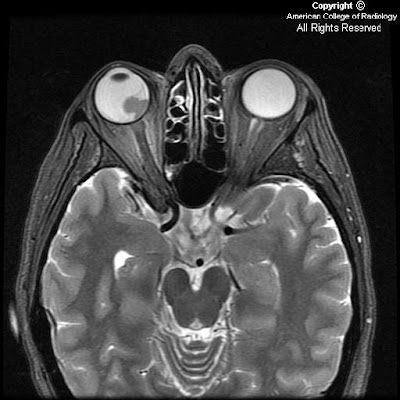
Thin section axial fat-suppressed fast spin-echo T2-weighted imaging
Figure 1 and Figure 2 show a V-shaped retinal detachment pointing toward the optic nerve. There is increased T1 signal posterior to the detachment suggesting hemorrhage.

Thin section axial fat-suppressed T1-weighted imaging
Figure 3 and Figure 4 show a mass arising from the nasal aspect of the right ocular globe. There is a collar button configuration to this mass, which assumes the typical configuration strongly suggesting elevation of Bruch’s membrane. The mass is near isointense to muscle on precontrast T1 and T2 weighted images. This mass extends towards the vitreous but no extension beyond the sclera is identified.

Postcontrast fat-suppressed T1-weighted imaging
Figure 5 and Figure 6: The mass is near isointense to muscle on pre-contrast T1 and T2 weighted images and demonstrates prominent contrast enhancement.


Diagnosis: Malignant melanoma of the uvea


Uveal melanoma is the most frequent primary intraocular malignant tumor in adults. It is rare in children. Its importance is that it is the main intraocular disease that can be fatal in adults.
Uveal melanoma can erupt through Bruch's Membrane. When this occurs, they develop a characteristic collar button configuration that extends through the vitreous chamber.
Uveal melanomas have differing MR characteristics, depending on the amount of melanin, which has paramagnetic properties. Melanomas containing a lot of melanin will demonstrate T1 hyperintensity, and markedly decreased T2 signal. However, in amelanotic or slightly melanotic melanomas, the typical MR pattern is isointense on T1, and slightly hypointense on T2.

Uveal melanoma is the most frequent form of intraocular malignancy in adulthood (4). As the uvea is the most vascular region of the globe, it is a common site for primary and metastatic neoplasm. Uveal melanomas comprise 70% of malignant intraocular tumors. It affects approximately 5-7 out of 1,000,000 people (3). Uveal melanoma can occur in any of the three subdivisions of the uvea: the iris, ciliary body, and choroid.

Uveal melanomas start with a flat growth profile along the choroid. With progression, they become elevated, and frequently extend through Bruch's membrane, where they can track into the subretinal space. With spread through Bruch's membrane, the melanoma can have a "mushroom shape" or "collar button" appearance that extends through the vitreous chamber.

Uveal melanomas typically appear as a solid, well-defined mass on magnetic resonance imaging. Melanin is paramagnetic, so in melanomas containing a lot of melanin, there is increased T1 signal with markedly decreased T2 signal. This signal intensity pattern is pathognomonic for uveal melanoma, as there are no other intraocular lesions with this appearance. In low-melanin or amelanotic melanomas, Magnetic Resonance imaging is less specific, but typically shows isointense signal on T1-weight images and slightly hypointense signal on T2-weighted images. Uveal melanomas typically have moderate to strong contrast enhancement following administration of gadolinium.

B-mode ultrasound typically shows a rounded, hypoechoic, highly vascular lesion. Retinal elevation and vitreous hemorrhage can also be seen, as these are complications of uveal melanomas. Uveal melanomas on unenhanced computed tomography appear sharply marginated, hyperattenuating, and elevated.

Uveal melanomas also have a propensity to metastasize hematogenously, and do so most frequently to the liver. Uveal melanoma is the most common fatal intraocular disease in the adult population.

Optimal treatment for uveal melanomas is controversial and clinical trials are ongoing. Large melanomas, typically greater than 10-mm in thickness, are usually managed with enucleation. For medium sized melanomas, 3-mm to 10-mm thick, plaque brachytherapy and external-beam radiation therapy have been accepted as alternatives to enucleation. For small lesions, less than 3-mm, routine monitoring with ultrasound is recommended as these may represent benign choroid nevi. These small lesions may also be biopsied, with a positive result placing the small melanomas into the medium melanoma treatment category.

Prognosis is dependent on many factors. Increasing tumor pigmentation is associated with a less favorable prognosis. Additionally, increasing size, infiltration through Bruch's membrane, and retinal detachment are all associated with a poorer prognosis. In metastatic disease to the liver, the mean survival has been reported to be nine months.